Saturday, March 31, 2012

Creating shocking memories


How does our brain make memories?  We know that a brain region called the hippocampus is important for forming new memories.  If this region is damaged or inhibited in mice or humans, memory formation is impaired.   

It has been hypothesized recently that a relatively small population of hippocampal neurons becomes activated during learning and that they act as a form of memory storage.  During memory retrieval these same neurons will become reactivated in such a way that the organism will “remember” the original stimulus.  If there were some way to tag these neurons during memory formation and then artificially reactivate them, you could test if these neurons are indeed acting as memory storage.

Two papers came out recently that used the same technique to tag specific neurons associated with a particular memory.  One paper by Garner et al published in Science, investigated if an artificially stimulated memory would impair or be incorporated into a naturally stimulated memory.  The other paper was rushed out by Nature the next week, probably because they realized the two papers are fairly similar and they didn’t want to be scooped.  The Nature paper, by Liu et al., asked if their artificially stimulated memory could induce a behavioral response on its own.  In other words, if you stimulate a certain set of hippocampal neurons associated with a memory, will the mouse act as if it’s really remembering the original stimulus?

Both papers used mice, which learned to associate a particular cage with a negative stimulus.  When mice are put into a cage, they usually walk around and aren’t too afraid.  However, if the base of this cage is electrified and the researchers give short electric shocks to the mice, they start to form a memory that this cage is a mini torture chamber.  (I use the term "torture" facetiously; I am sure this research was approved by the universities to make sure no animals were ever in serious pain.)  If they put the mice back into a normal cage, the mice recover and go on about their business.  The next day, the mice are put back into the torture cage and they recognize how it’s decorated and remember that they’ll get shocked.  The mice are afraid and freeze in anticipation of being shocked.  This is called “fear conditioning” and is a way of testing if mice have learned this association and can remember it days later.  The amount of freezing is measured as the behavioral output of memory formation and recall.

Memory formations of different cage decorations activate different sets of neurons in the hippocampus.  The researchers wanted some way of tagging these particular neurons so they could be activated at a later date using an artificial stimulus.  Okay, what follows is a complicated molecular biology scheme, but I think it’s really ingenious.

Artificial activation of hippocampal neurons
I will describe how Liu et al. were able to artificially activate particular hippocampal neurons, but the strategy used by Garner et al. is very similar.  When mice are placed into a new cage, they notice the color of the walls, the shape of the cage, etc.  This activates a certain subset of neurons in the hippocampus.  When neurons are highly activated, they start expressing a gene that makes a protein called c-Fos.  This protein is what we call a transcription factor, which can activate expression of other genes.  Normally, it would activate genes associated with making the neuron more easily activatable for the future.  This is what we call “cellular memory”.  The neurons “remember” being activated once and will be more likely to fire in the future. 

The mice used in these experiments were also expressing some genes that were made by the researchers and put into the mice.   During memory formation, C-Fos binds all its normal target genes, but it also binds a construct made by the researchers; it activates expression of the tTA gene, which makes another transcription factor.  tTA activates expression of another gene called Channelrhodopsin (ChR2).  This gene makes an ion channel that opens in the presence of blue light.  When it opens, it lets positive sodium ions into the neuron, which activates the cell.  Feeding the mice a chemical called doxycycline can inhibit the action of tTA.


The left vertical pathway is what happens normally during memory formation.  The horizontal pathway is what happened, in addition, in the transgenic mice used by Liu et al.

That’s complicated, but here’s the important part:
1) Neurons that are activated when the mouse is in a new cage will make C-Fos and turn on this molecular system.
2) The activated neurons will end up expressing a light-activated channel (ChR2).
3) Researchers can then turn on these specific neurons with light whenever they choose.  A neuron that was not originally activated by the new cage will not have the light activated channels.
4) Researchers can thus label active cells and turn them on or off with light.  This whole expression system can also be turned off with doxycycline, so neurons can be labeled only during the experimental time period.

In the other paper, instead of expressing ChR2, the mice expressed a different receptor that can be activated with another chemical called CNO.  Chemical activation isn’t as fast as light activation, so this was more for long-term activation (hours vs. seconds).

The Experiments
Liu et al. wanted to see if they could induce the freezing behavior of mice, just by activating the subset of neurons with light.  Here’s the set-up: the mice were put in Cage A and given electrical shocks.  If they were put back in Cage A, they would freak out and freeze.  If they were instead put in Cage B, which looked different, they were not afraid and did not freeze.  Remember that these mice also were expressing ChR2 in the neurons that had been originally activated by Cage A.  When the mice were in neutral Cage B, the researchers shined blue light onto their hippocampus (via an implanted fiber optic cable).  The blue light activated ChR2, activated the neurons associated with Cage A, and the mice froze!  The neurons that were activated by the Cage A context had formed an association with fear in the brain, so when they were activated artificially later, the mice became afraid.  Wow!  Artificially induced memory recall.

The experiments done by Garner et al. were interesting too, but a bit more complicated.  They put the mice in Cage A without an electric shock and labeled these neurons with their receptor.  Then they moved these mice to Cage B and gave them an electric shock.  So the mice were fear conditioned for Cage B, but their neurons for Cage A could still be artificially activated by the researchers.  Next they put the mice in Cage B again and they were scared and froze.  Then they artificially activated the Cage A neurons (by giving them the CNO activator) while the mice were in Cage B.  This “memory” of the neutral Cage A was able to override the fear memory of Cage B and the mice did not freeze anymore.  In other words, the internal retrieval of a neutral memory interfered with the mouse’s fear memory.  In some distant future, a similar technique potentially could be used by humans trying to face a fear, but that’s pure speculation.

In another experiment done by Garner et al. (see figure below), they put the mice in neutral Cage A and labeled these neurons.  Then they did fear conditioning in Cage B, but this time they also artificially activated the neurons for Cage A at the same time.  At this point neurons for Cage B were naturally being activated, while the Cage A neurons (which are different) were also active.  One day later, when they tested the memory of the mice in Cage B, the mice no longer froze; they weren’t afraid of Cage B.  But if they were in Cage B and the Cage A neurons were activated by the drug CNO at the same time, the mice froze.  So what’s going on with this?  During fear conditioning, the mice were seeing Cage B but were also having Cage A neurons activated.  The fear memory incorporated both Cage A and Cage B neurons into the memory trace in the brain.  Wow!  They created a synthetic hybrid memory.   This implies that if you’re thinking about one thing while a memory of something else is being formed, the two things may merge together into some other non-realistic memory.  In the words of the authors “the internal dynamics of the brain at the time of learning contribute to memory encoding”.

Formation of a hybrid memory.  Neither Cage B or Cage A alone can induce the fear memory.  The mouse needs both sets of neurons to be activated at the same time to remember the electric shock from Cage B.  Neurons for Cage A are activated artificially with the drug CNO.  Neurons for Cage B are activated naturally by the cage itself.

Conclusions
I love both of these papers.  They used new techniques in molecular biology to really address new questions about how our brain encodes memories.  Both demonstrated that a discrete population of neurons in the hippocampus is responsible for forming new spatial memories (about the visual nature of the cages).  Liu et al. showed that they could elicit an output of a memory just by reactivating the neurons that had been active during learning.  Garner et al. demonstrated that the internal self-generated activity of neurons in the brain can have an impact on memory formation.  Kudos to both groups and there’s no need to feel scooped, Authors, because the two papers are complimentary in the experimental questions that were answered.

Sunday, March 25, 2012

Hot times are a-bloomin'


Remember a month ago, before the rains started, when it was warm and sunny and all the trees in Berkeley were blooming?  The Magnolias and plum trees were particularly beautiful. 

From Berkeleyside blog

How do the plants know that it’s warm and time to bloom?  Plants can sense day length and will begin to grow and bloom when the length of sunlight increases in spring.  Our trees were blooming in January and February, though, so plants must also be able to sense temperature changes too.  This week’s paper addresses the molecular changes that occur to induce flower growth in response to warmer temperatures, even when the daylight is still reduced in winter.  The paper by Kumar et al. was published online in Nature.

The transcription factor PIF4
The authors focused their studies on a protein called PIF4.  This protein binds to DNA and helps direct gene expression (or transcription), which makes it a transcription factor.  In normal, wildtype Arabidopsis plants, there is early flowering at warm temperatures, but in mutants that do not have PIF4, flowering is delayed.  In contrast, when PIF4 is overexpressed, early flowering is induced.  These results show that flowering at warm temperatures depends on PIF4.

The authors go on to show that PIF4 binds DNA just upstream of a gene called FT, which is necessary for flowering.  One can imagine a simple pathway where higher temperatures induce PIF4 to bind to the FT gene and activate gene expression.  Increased FT then initiates cellular pathways that signal the plant to go into bloom.

How do increased temperatures increase PIF4 binding to the FT gene?
1) Maybe high temperatures increase transcription of PIF4.  The authors measure RNA levels of PIF4 at different temperatures and find that there is more expression at higher temperatures, but not enough to account for the extreme differences in flowering.
2) Maybe high temperatures make the protein more stable.  In this case you would expect to see less PIF4 protein at cold temperatures, but the authors find that protein levels are unchanged by temperature.
3) Do high temperatures help the binding interaction between PIF4 and FT?  Yes, there is more PIF4 bound to FT at higher temperatures, but why?

Nucleosome H2A.Z
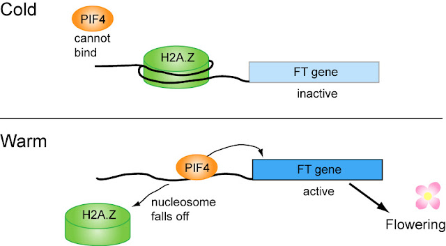
In past posts, I’ve talked about histones in the context of epigenetics.  DNA wraps around histone proteins to help pack the DNA into a smaller package.  The collection of histones that DNA wraps around is called a nucleosome.  When DNA is wrapped around nucleosomes, other proteins cannot bind to the DNA and gene expression doesn’t happen.  There is a nucleosome called H2A.Z that binds near the FT gene in the same place where PIF4 needs to bind.  Therefore, when the nucleosome is present, PIF4 cannot bind and activate expression of FT and there will be no flowering.  H2A.Z binding to FT is temperature dependent in the exact way you would expect.  In cool temperatures, H2A.Z binds FT DNA, so PIF4 cannot bind and FT will not be expressed.  In warm temperatures, for whatever reason, the H2A.Z nucleosome comes off of the DNA, allowing PIF4 to bind.
 
Adapted from Figure 4C of Kumar et al., 2012, Nature.

It’s nice when there is a relatively simple molecular explanation for something we observe in the environment all the time.  This subject is also important because global warming will continue to induce earlier and earlier flower blooming, which could be detrimental to food crops.  

Sunday, March 18, 2012

The rewards of sex and alcohol


There’s a new fruit fly article making the rounds this week.  The public loves a good fruit-flies-acting-like-us story.  The take home message is that male fruit flies that are deprived of sex are more attracted to alcohol (which affects them in a way similar to humans).  Fruit flies like sex!  Fruit flies like alcohol!  Fruit flies are just like us!  Besides the fact that this is funny, who cares, right?  Well, these findings have important implications for the study of addiction.  Let’s dive into the science of the article to understand the connections to addiction.

Sexual deprivation and alcohol intake
The first thing the authors did was establish a behavioral test, which measured attraction towards alcohol.  Flies were put into a jar and in the lid were little tubes filled with fly food (sugar and yeast) or filled with fly food spiked with 15% ethanol.  The flies could choose to eat as much as they wanted of either type of food.  The researchers could measure how much of the food was consumed and create a ratio, which they called the preference index.  The more ethanol-spiked food was consumed, the higher the number for the preference index (in other words, the flies prefer ethanol consumption).  It’s previously been shown that flies will consume more of the ethanol-spiked food, even if the food tastes bad to them.

Next the authors tested two different sets of males.  In one set, the males were put into a container with many virgin females and allowed to mate for 6 hours (!)  In the other set, individual males were introduced to females who had already mated and were, therefore, not receptive.  In other words, these males never mated and were rejected day after day.  These two groups of males were then put in the jar with the food and ethanol and allowed to choose what to consume.  The males who were rejected consumed much more ethanol containing food, whereas the males who had mated ate the normal food.   (The New York Times has a nice video of flies mating or being rejecting, which is associated with their article about this research.)

Is this like drowning your frustrations in a beer?  Sort of.  The males who had been rejected, experienced two negative things: (1) They tried to mate and were rejected. (2) They didn’t have sex.  Is it the rejection or the lack of sex that drives them to drink?   

The authors tested this by putting males in with decapitated virgin females.  Yes, that’s right—these females were dead and lacked heads.  Males will still try to mate with these females, who obviously can’t reciprocate.  However, since the females are dead, they do not actively reject the males.  These males who couldn’t mate also showed a preference for ethanol, so it’s not the “anger” of being rejected that drives them to ethanol, it’s just not having sex.

Neuropeptide F
The authors measured levels of neuropeptide F (NPF) in their two sets of male flies, and found that males who had not mated had lower levels of NPF RNA and protein.  What is NPF?  Neurons communicate with each other by using chemical signals.  Some of these signals are small proteins, called neuropeptides, which act as modulators of neuronal activity and are often involved in behaviors.  The authors wondered if they could control the flies’ attraction to ethanol by experimentally altering the NPF levels.  Males who had no sex had lower NPF and higher preference for ethanol.  Therefore, one might expect that a mated male who normally does not want ethanol, could develop a preference for it, if his NPF concentration was artificially decreased.  


The authors decrease NPF levels by expressing inhibitory RNA (RNAi) for that gene.  RNAi is a way of decreasing expression of a gene of interest, without actually mutating the gene.  Mated males who expressed RNAi for NPF (their NPF levels were decreased), had a greater preference for ethanol than the controls. 

In the complimentary experiment virgin males were used who have not had sex, have decreased NPF levels and a greater preference for ethanol.  In order to change their ethanol preference, the authors would need to somehow artificially increase NPF levels.  Instead of doing this directly, what they did was force the neurons that would be activated by NPF into a state where they were always activated regardless of NPF levels.  In other words, they bypassed the need for NPF and just activated the output cell.  Virgin males with their cells artificially activated (as if there were a lot of NPF around) lost their preference for ethanol.


To summarize, mating increases NPF expression and increased NPF expression makes the flies less attracted to ethanol. 

The authors go on to show that mating is rewarding to flies, as is ethanol consumption and having high levels of NPF.  The NPF signal in the brain acts as a “reward” center.  When fruit flies have sex, this area of the brain is activated and they feel “good” and “satisfied”.  However, when the fly is unable to have sex, the reward center is under-activated and the fly fills in this lack of signals by consuming alcohol.  Ethanol, as well as other drugs, also activates this reward center and brings the signaling back to normal levels.  The flies which had sex don’t need ethanol, because their reward center has already been activated sufficiently.  Drug abuse then might be viewed as the brain trying to get that reward signal, which for whatever reason is not being satisfied by natural stimulants such as food, sex or other social interactions.

Wednesday, March 14, 2012

The buzz about novelty-seeking


One topic I have always been interested in is how complex behaviors are driven by genetics and molecular biology.  For instance, all the courtship rituals of a male fruit fly are basically encoded by a single gene, and more specifically one small exon (a chunk of DNA within a gene).  A recent paper by Liang et al. which was published in Science, looked at novelty-seeking behavior in honeybees.  The nice thing about this paper is that they started out by finding bees that like to scout for food and nests and then worked backwards to figure out what is different about these bees compared to their boring, steady-as-she-goes sisters.

Novelty-seeking in bees
 There are two types of novelty-seeking behaviors which the authors investigate. 

1) Food scouting: Even when there are plentiful known food sources, some bees (5-25% of foragers) still go out to look for new sources.  The other foraging bees rely on these food scouts, who tell them where the new food source is.  The food scouts are obviously important for a colony, so the bees always know where to get food.
Photo taken by Rebecca Wenk 2010.

2) Nest scouting: When a swarm has left its colony in search of a new location, a few bees (<5%) will search out for the best place to build a new hive.  They will tell the rest where to go and lead them there.

Are nest scouts the same bees as food scouts?

The researchers identified nest scouts in artificial and natural swarms and marked them.  Then they waited to see if these nest scouts would become food scouts as well.  They moved the hives every night to a new location.  The bees that seek new food sources will not be thrown by the new environment and will be able to find new food sources quickly.  Under these circumstances, the authors found there was a trend for the former nest scouts to also be food scouts, though it wasn’t a hard-and-fast rule.

Molecular underpinnings
All worker bees are female and have the same genetic make up— they’re all identical sisters.  What would make some bees more likely to explore novel environments?  To examine this question, the authors collected food scouts.  While the bees slept in their hives, the authors introduced a new feeder in different locations each day.  They collected bees that visited the new feeder twice, when it was in two different locations.  Once they had the food scouts, they performed a microarray to determine differences in gene expression between food scouts and non-scout controls.

If you want to know more about how microarrays work, visit my methods section.  All you really need to know is that a microarray looks at every single gene and determines how much it is expressed.  Since all bees have the exact same genes, the only differences are going to arise by how these genes are expressed (remember: not all genes are expressed at a given time).

The authors found that scouts have differential expression of various neurotransmitter systems.  Neurotransmitters are the signals that are sent from one neuron to another.  They found that scouts had higher expression of genes involved in GABA and glutamate neurotransmission.  In general, GABA is used to inhibit neurons, and glutamate is the standard excitatory neurotransmitter.  GABA and glutamate may control different neuron circuits in the brain.  They also saw a decrease in a receptor for dopamine.  What this implies is that when the brain has increased GABA and glutamate signaling, but decreased dopamine, it makes the bees want to seek out novel environments (or maybe it makes them less afraid to try something new). 


To prove that this hypothesis is correct, the authors took some non-scouts and fed them glutamate.  These non-scouts with extra glutamate were more likely to search for new food.  In other words, they switched the behavior of non-scouts to novelty-seeking by changing the balance of neurotransmitters in their brain.  Unfortunately, this didn’t work when they tried to do the same thing with dopamine.  They inhibited the dopamine receptor, which should also increase food scouting, but it actually made the non-scouts even less likely to scout for food.

It’s cool that the authors did behavioral studies in bees, but I feel like something was missing in this paper.  Okay, neurotransmitter systems are expressed differently, but why?  A paper from last year showed that queen bees have different epigenetic marks than worker bees, so maybe this is what’s going on with the novelty seekers.  Is there some difference in their environment as they develop, which causes epigenetic changes and differential gene expression?  There is a lot more to explore in this topic.

Thursday, March 8, 2012

Translation of Touch


The language of neurons is ion movement across the cell membrane.  When a neuron becomes activated, positive sodium ions rush into the cell through ion channels (gates in the cell membrane that can open and close).  This flow of ions (also called “current”) is propagated from cell to cell, to initiate some sort of response (a movement, a thought, etc).

How are the five senses translated into ion flow?  For olfaction (and taste), odor molecules bind to specific receptors which activate a signaling cascade in the cell which eventually leads to ion channels opening.  When a sound wave enters our ear, it moves little hairs on the auditory sensory cells, which cause ion channels to open.  The sense of touch works in a similar way: there are receptors, called mechanoreceptors, which are gated by physical pressure to the cell membrane (Figure 1).  It’s analogous to pushing against a swinging door; with enough pressure, the door will open up and let things pass into the building.


In this week’s paper, which appeared recently in Nature, Kim et al. characterize a new mechanoreceptor called Piezo ("piezo" comes from the Greek meaning “to press”).  Mechanoreceptors have been studied before in Drosophila fruit flies, but Piezo is the first one that is evolutionarily conserved in mammals too. 

Piezo mutants
The authors made a mutant fly line that is missing the piezo gene.  These flies survive and seem healthy enough, but when they tested the larvae (the “maggots” if you will), the authors discovered that they had lost their response to strong touch. 

To do these experiments, they used what’s called a von Frey filament.  These are little hairs made from varying diameters of nylon, which they pressed against the larvae until the filaments bent.  The amount of force needed to bend the filament varies with the diameter of the filament, so you always know how much force was applied to the filament and thus to the larva.  Larvae don’t like being touched (it might be equivalent to someone punching us), so they will respond by rolling or wriggling away.

Wildtype larvae (not mutated) responded to the strong touch more than 80% of the time, but the piezo mutants responded only 34% of the time.  Interestingly, though, the mutants responded normally to both high temperature and gentle touch, which shows that some other receptor must transduce these sensory signals.

Where is Piezo expressed?
Since Piezo is necessary for noxious (i.e. strong) mechanosensation, it must be present in sensory neurons.  Drosophila larvae have different types of sensory neurons lining their body walls.  One subset of these sensory neurons is called ppk-positive, because these cells express another mechanoreceptor called ppk (short for pickpocket).  The authors find that every ppk-positive cell also contains Piezo.  In their first experiment, they used mutant flies where every single cell was missing the piezo gene.  Now the researchers knocked down expression of piezo only in ppk-positive cells, which caused the same decrease in number of larvae that responded to strong touch.  They were also able to rescue the mutant piezo flies by expressing the normal Piezo protein only in ppk-positive cells.  In other words, Piezo and the other mechanoreceptor, PPK, act in the same sensory neurons.

Piezo and PPK both mediate mechanosensation
Recall that the piezo mutants had a reduced response to noxious stimuli, but the response wasn’t completely abolished.  What accounts for the 34% response rate?  Could it be PPK (which is still present in the piezo mutants)?  Sure enough, when Kim et al. reduced expression of both piezo and ppk, the response to strong touch was effectively abolished (see Figure 2 below). 


This means that the sensory neurons express two different mechanoreceptors that both respond to strong pressure by passing positive ions into the cell.  Larvae definitely want to get away from painful stimuli, so it makes sense that there would be redundant pathways.  It’s such a simple and elegant way to sense touch.  Mechanoreceptors act as the translators between a physical stimulus (touch) and the electrical signaling of neurons.


Saturday, March 3, 2012

Take two HDAC inhibitors and call me in the morning

Changes in the environment, either at the level of cells, or on an organismal scale, can activate enzymes that chemically modify the DNA or DNA-associated proteins in such a way that gene expression is altered. For instance, it’s been shown that parental neglect of young mice, causes a number of their genes to be turned off via epigenetics. In this week’s paper, published online in Nature, the authors make a link between Alzheimer’s Disease, epigenetics and widespread decreased expression of genes that are important for cognitive function and learning. 

Histone Acetylation
The first thing that you need to understand is the connection between histone acetylation and gene expression.

Question 1: What is a histone?
We have 46 chromosomes in every cell and each chromosome is made up of a very, very, very long strand of DNA. Think about what happens to your electrical cords and chargers and headphones when you throw them all into a drawer: all the wires get wrapped up together and twisted into knots. That could happen to our long strands of DNA, which would be a disaster. Luckily, our cells are super organized and wrap sections of DNA around spools of histone proteins. This is sort of like the tabs on a Mac charger, around which you can wrap the extra cord. The DNA wraps two times around each core of histones and then spans to the next histone and wraps around two times, etc. The histones help keep the DNA organized, but they also act as a way to control DNA expression.


Question 2: What is acetylation?
An acetyl group is a small chemical group (-COCH3) that can be added to larger molecules by enzymes. The histone proteins have tails that hang out and which can be acetylated in a dynamic process.

Question 3: What does this have to do with gene expression?
If the DNA wraps around the histones very tightly, the protein machinery that expresses DNA cannot get access to the DNA and gene expression is turned off. This is one way that histones can control DNA expression. When histones become acetylated, the DNA loosens its grip on the histones, so there is plenty of room for proteins to bind DNA and gene expression is turned on.

+ Acetyl = active genes
- Acetyl = inactive genes

Question 4: What adds acetyls to histones?
Enzymes called Histone Acetyl Transferases (HAT) add acetyl groups, and enzymes called Histone Deacetylases (HDAC) take off acetyls. If you have a high level of HDACs around, they will take off lots of acetyls, DNA will wrap tightly around histones and gene expression will be turned off (see top part of figure below. Figure from Pons et al., 2009, European Heart J)

Increased HDAC --> - Acetyl --> tightly wrapped DNA --> inactive genes


HDAC in Alzheimer’s model
The authors investigated the role histone acetylation may play during the progression of neurodegeration associated with Alzheimer’s Disease (AD). A major aspect of AD is the accumulation of protein aggregates in the brain made up of amyloid beta (amyloid plaques). There are mouse models of AD which have these amyloid plaques, neurodegeneration (their brain cells die) and cognitive defects. The authors measured levels of different HDACs in one of the AD mouse models and found that HDAC2 was greatly overexpressed in the brain compared to control mice. As would be expected, histone acetylation was reduced (remember they take off acetyl groups from histones). They next looked at the expression levels of genes that have been implicated in the process of learning and memory. When we learn something, certain genes are activated in nerve cells, which can lead to long lasting changes to the functioning of the cells (maybe they will become easier to activate or maybe the cell will make more synaptic connections with other nerves). Gene expression was reduced, which may be one reason for the cognitive defects associated with AD.

Restoring cognition
To summarize, there is more HDAC2 around in AD mice, so there is less histone acetylation and gene expression is decreased. The authors reasoned that perhaps they could restore the ability of these mice to learn and remember if they could stop the increase in HDAC2. In mice and other experimental organisms, you can lower expression of any gene of interest by expressing what’s called inhibitory RNA (RNAi) for that gene. This is basically the complementary sequence of that gene (which is DNA) in the RNA format. What’s important here is that the authors made a RNAi for HDAC2 and stuck it in a virus which was used to get this molecule into the mouse brain. The RNAi did its job and brought the levels of HDAC2 in AD mice back down to a normal level. Expression of the “learning” genes was restored and their neurons made more synaptic connections than the AD mice with high HDAC2. Interestingly, the RNAi wasn’t able to reverse the neurodegeneration; once the neurons die in AD mice, they are dead forever. However, the fact that their remaining neurons made more synaptic connections was a good sign that learning could happen again. The authors set up a series of different learning tasks for the mice, and the RNAi mice with normal levels of HDAC2 performed as well as the controls, whereas the AD mice with high HDAC2 underperformed.

Being able to rescue cognitive defects in AD mice by simply altering the levels of a histone deacetylase is pretty amazing. It shows that the AD mice brains are still capable of learning and of growing more synapses, but they are constrained by the gene expression block imposed by HDAC2. This would be a good direction to go when investigating new treatments for Alzheimer’s Disease. In fact, the authors looked at tissue from human brains and found that patients who died from AD had increased HDAC2 protein expression compared to healthy controls. This is a good indication that what worked in the mouse model could also work in humans.


Besides this, the authors did a whole set of experiments to show what causes the HDAC2 overexpression in the first place. Remember that one of the main causes of AD is protein aggregation, or amyloid plaques. These big globs of proteins stress out the cells, which may activate different protein pathways to try to deal with the situation. One of the proteins that is turned on is called GR1 which activates expression of the HDAC2 gene. Basically, the cell is freaking out and tries to help, but HDAC2 overexpression is actually bad and prevents neurons from learning.

Stressed cell --> GR1 --> HDAC2 gene expression --> less histone acetylation --> compact DNA --> inactive genes --> inability of cell to grow synapses --> no learning

This paper was ridiculously thorough. Whereas most papers would have stopped at restoring cognition (this has big implications), the authors kept going on to describe the whole signaling pathway. And to think, it all has to do with epigenetics and little acetyl groups of histones. Amazing!